一、本期重点:
doi: 10.1364/BOE.6.003564
published:2015.08.25
内容介绍:
老年性黄斑病变(AMD)是发展中国家年龄大于50周岁人失明的主导因素。AMD的一种特征是存在脉络膜新血管化(CNV),即来自脉络膜的病变的新血管通过布鲁赫膜的堵塞生长至无血管的外层视网膜。光学相干层析造影术是一项非侵入性的、深度分辨的三维成像技术,广泛应用于视网膜形态学临床和科研。近年来,该项技术被用于实现对患有AMD疾病的病人的脉络膜新生血管的成像。
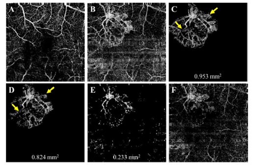
对CNV的识别与量化在AMD疾病的临床诊断和治疗中具有重要的价值。该文报道了一种自动的CNV区域探测识别算法。依靠降噪和一个突出的探测模型攻克了CNV中的伪影和不均匀问题。该算法使用的模型基于强度、方向和位置信息,能够识别CNV清晰的轮廓。实验结果和对CNV的手动描述结果一致。

图一:某AMD患者CNV区域的OCT微血管造影图像。未做额外算法处理的(A)内视网膜血管图和(B)外视网膜血管图;(C)专家人为描绘的和(D)采用自动特征算法得到的CNV区域图像;(E)采用先前的自动算法;(F)未做额外处理的脉络膜血管层造影图。
doi: 10.1364/BOE.7.000816
published:2016.02.09
内容介绍:
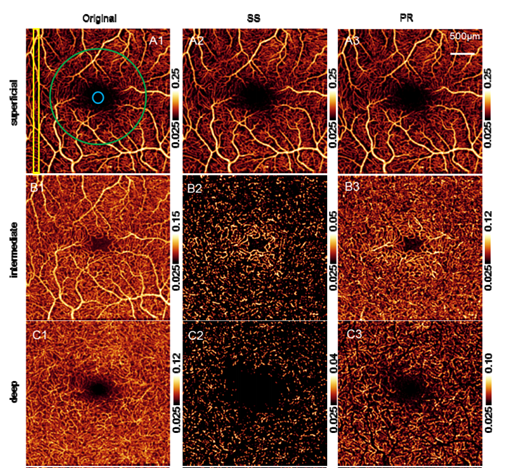
在光学相干层析造影(OCT-A)中,浅层血管的投射伪影严重影响深层血管网络的清晰成像识别。该小组提出了一种新颖的算法,通过分辨原位置的和被投影的血流信号消除伪影。由于实际血管位置处的信号强度归一化后的解相关值比同一轴向扫描线内的所有伪影血管的解相关值要高,基于以此该算法能够识别实际血流位置处的血管。这种投影分辨的算法有效地抑制微血管造影图中的投射伪影。

图一:人眼视网膜OCT-A图像。(A1-C1)未做投影抑制;(A2-C2)采用标准的相减(SS)法抑制投影后;(A3-C3)采用该文提出的投影分辨算法抑制投影。A行:浅表血管;B行:中间层血管;C行:深层血管。
二、简讯:
doi: 10.1364/OL.41.001058
published:2016.03.01
Abstract:
We proposed a single-shot spatial angular compounded opticalcoherence tomography angiography (AC-Angio-OCT) for blood flow contrast enhancement. By encoding incidentangles in B-scan modulation frequencies and splitting themodulation spectrum in the spatial frequency domain, angle-resolved independent subangiograms were obtainedand compounded to improve the flow contrast. A full spaceof the spatial frequency domain allows a wide modulationspectrum. To get access to the full space of the spatial frequencydomain and avoid the complex-conjugate ambiguityof the modulation spectrum, a complex-valued OCT spectralinterferogram was retrieved by removing one of theconjugate terms in the depth space. To validate the proposedconcept, both flow phantom and live animal experimentswere performed. The proposed AC-Angio-OCToffers a ∼50% decrease of misclassification errors, and animproved flow contrast and vessel connectivity, which contributesto the interpretation of OCT angiograms.
doi: 10.1364/OL.41.003944
published:2016.08.19
Abstract:
The current temporal, wavelength, angular, and spatialaveraging approaches trade imaging time and resolutionfor multiple independent measurements that improve theflow contrast in optical coherence tomography angiography (OCTA). We find that these averaging approaches areequivalent in principle, offering almost the same flow contrastenhancement as the number of averages increases. Based on this finding, we propose a hybrid averaging strategyfor contrast enhancement by cost apportionment. Wedemonstrate that, compared with any individual approach, the hybrid averaging is able to offer a desired flow contrastwithout severe degradation of imaging time and resolution. Making use of the extended range of a VCSEL-basedswept-source OCT, an angular averaging approach by pathlength encoding is also demonstrated for flow contrastenhancement.
doi: 10.1097/IAE.0000000000000846
published:2015.11
Abstract:
To use optical coherence tomography (OCT) angiography to monitor the short-term blood flow changes in choroidal neovascularization (CNV) in response to treatment.In this retrospective report, a case of exudative CNV was followed closely with OCT angiography over three cycles of antiangiogenic treatment. Outer retinal flow index, CNV flow area and central macular retinal thickness were measured.Quantitative measurements of CNV flow area and flow index showed rapid shutdown of flow over the initial 2 weeks, followed by reappearance of CNV channel by the fourth week, preceding fluid reaccumulation at 6 weeks.Frequent OCT angiography reveals a previously unknown pattern of rapid shutdown and reappearance of CNV channels within treatment cycles. OCT angiographic changes precede fluid reaccumulation and could be useful as leading indicators of CNV activity that could guide treatment timing. Further studies using OCT angiography in short intervals between antiangiogenic treatments are needed.
doi: 10.1364/BOE.7.001905
published:2016.04.18
Abstract:
Recent advances in optical coherence tomography (OCT)-based angiography have demonstrated a variety of biomedical applications in the diagnosis and therapeutic monitoring of diseases with vascular involvement. While promising, its imaging field of view (FOV) is however still limited (typically less than 9 mm2), which somehow slows down its clinical acceptance. In this paper, we report a high-speed spectral-domain OCT operating at 1310 nm to enable wide FOV up to 750 mm2. Using optical microangiography (OMAG) algorithm, we are able to map vascular networks within living biological tissues. Thanks to 2,048 pixel-array line scan InGaAs camera operating at 147 kHz scan rate, the system delivers a ranging depth of ~7.5 mm and provides wide-field OCT-based angiography at a single data acquisition. We implement two imaging modes (i.e., wide-field mode and high-resolution mode) in the OCT system, which gives highly scalable FOV with flexible lateral resolution. We demonstrate scalable wide-field vascular imaging for multiple finger nail beds in human and whole brain in mice with skull left intact at a single 3D scan, promising new opportunities for wide-field OCT-based angiography for many clinical applications.
供稿:李培


