
一、本期重点:
doi: 10.1364/OL.40.005355
published:2015.11.10
内容介绍:
共焦显微术(confocal scanning microscop—CSM)是目前光学显微领域中应用的最广泛的显微技术。理论上共焦能在宽场显微的基础上提高1.4倍的分辨率,但实际上这只是在不考虑信噪比的情况下得到的。图像扫描显微术(Image scanning microscop—ISM)解决了这个问题,但是系统过于复杂。这篇文章使用一个四象限探测器代替以往ISM的阵列探测器,成功的简化了系统装置。通过仿真模拟,使用四象限探测器的ISM(QISM)可以达到ISM的分辨率,并且可以提升1.5倍的信噪比,同时并不牺牲光学切牌能力。
该技术主要应用了导波光学相关理论。图一是各向异性“漏模”空间光调制器的结构,铌酸锂基底上集成了了质子交换波导管,尾部安装声波换能器。多个波导管集成在一起,全息信息耦合在声波信号中,输出信号会发生相应变化。
由此制造的空间光调制器可以达到更高的带宽,更低的衍射角,高可扩展性等一系列优点,几乎可以解决现在的各方面困难。
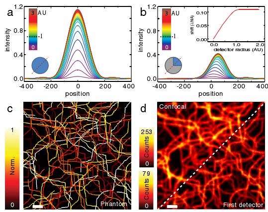
图一 模拟结果(a)小孔尺寸由0.1个艾里斑到3.0个艾里斑时的共焦PSF的强度。(b)小孔尺寸由0.1个艾里斑到3.0个艾里斑时的右上探测器的PSF强度。(c)模拟样品。(d)成像结果比较
2.表面一微米范围内牛精子游动跟踪(Two-dimensional slither swimming of sperm within a micrometer of a surface)【Nature Communications】
doi:10.1038//ncomms9703 | www.nature.com/naturecommunications
published:2015.11.10
内容介绍:
精子在表面的运动情况是受孕过程的重要步骤,而且这个活动目前为止还没有研究清楚。全内反射荧光显微方法能够观察表面1μm范围内的样品情况,在本篇论文中作者观察了活性人和牛的精子在表面的活动情况,根据观察结果在近表面区域,精子出去一种摆动式前进方式,这种方式与近表面和自由空间里的运动是很不同的,由于精子在不受限制的运动时,其鞭毛是螺旋的而且精子头部也是连续的转到前进。牛精子在表面运动比较慢,由于其鞭毛螺旋转幅度在近表面区域会变小,因而产生的对动力也会很小。相对比的是,人的精子能快50%,这意味着人类精子更适合高粘粘性和狭窄的人类输卵管。
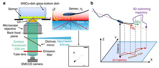
图二 全内反射荧光显微系统观察近表面精子。(a)结构示意图;(b)用于运动描述的笛卡尔坐标系
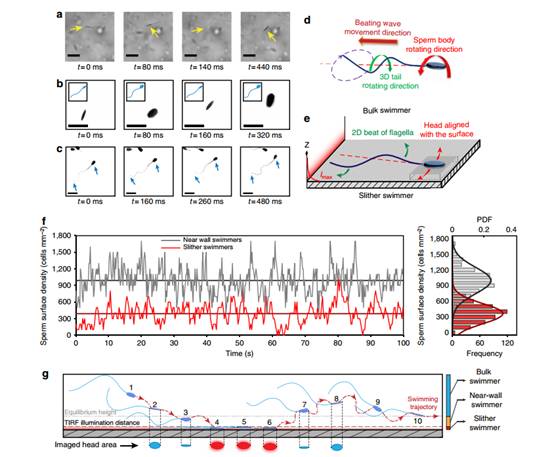
图三 自由空间运动与近场摆动运动的对比。a、宽场图像;b、荧光图像;c、全内反射图像;d、自由空间螺旋运动;e、近场摆动运动;f、精子头部在运动过程中的在像平面上的成像强度时间分布;g、精子运动示意图。
二、简讯:
1.使用on-chip和自组装微透镜实现宽敞纳米粒子探测(Wide-field optical detection of nanoparticles using on-chip microscopy and self-assembled nanolenses)
doi:10.1038/ NPHOTON.2012.337
published:2013.1.20
Abstract:
The direct observation of nanoscale objects is a challenging task for optical microscopy because the scattering from anindividual nanoparticle is typically weak at optical wavelengths. Electron microscopy therefore remains one of the goldstandard visualization methods for nanoparticles, despite its high cost, limited throughput and restricted field-of-view.Here, we describe a high-throughput, on-chip detection scheme that uses biocompatible wetting films to self-assembleaspheric liquid nanolenses around individual nanoparticles to enhance the contrast between the scattered and backgroundlight. We model the effect of the nanolens as a spatial phase mask centred on the particle and show that the holographicdiffraction pattern of this effective phase mask allows detection of sub-100 nm particles across a large field-of-view of>20 mm2. As a proof-of-concept demonstration, we report on-chip detection of individual polystyrene nanoparticles,adenoviruses and influenza A (H1N1) viral particles.
2.使用虚拟探测实现快速超分辨线扫描显微(Rapid super-resolution line-scanning microscopy through virtually structured detection)
doi: 10.1364/ OL.40.001683
published:2015.4.15
Abstract:
Virtually structured detection (VSD) has been demonstrated to break the diffraction limit in scanning laser micros-copy (SLM). VSD provides an easy, low-cost, and phase-artifact-free strategy to achieve super-resolution imaging.However, practical application of this method is challenging due to a limited image acquisition speed. We reporthere the combination of VSD and line-scanning microscopy (LSM) to improve the image acquisition speed. A mo-torized dove prism was used to achieve automatic control of four-angle (i.e., 0°, 45°, 90°, and 135°) scanning, thusensuring isotropic resolution improvement. Both an optical resolution target and a living frog eyecup were used toverify resolution enhancement.
doi:10.1038/nphoton.2015.216
published:2015.2.16
Abstract:
Medical X-ray imaging requires cost-effective and high-resolution flat-panel detectors for the energy range between 20 and 120 keV. Solution-processed photodetectors provide the opportunity to fabricate detectors with a large active area at low cost. Here, we present a disruptive approach that improves the resolution of such detectors by incorporating terbium-doped gadolinium oxysulfide scintillator particles into an organic photodetector matrix. The X-ray induced light emission from the scintillators is absorbed within hundreds of nanometres, which is negligible compared with the pixel size. Hence, optical crosstalk, a limiting factor in the resolution of scintillator-based X-ray detectors, is minimized. The concept is validated with a 256 × 256 pixel detector with a resolution of 4.75 lp mm−1 at a MTF = 0.2, significantly better than previous stacked scintillator-based flat-panel detectors. We achieved a resolution that proves the feasibility of solution-based detectors in medical applications. Time-resolved electrical characterization showed enhanced charge carrier mobility with increased scintillator filling, which is explained by morphological changes.
doi:10.1038/nphoton.2015.223
published:2015.2.23
Abstract:
Rapid progress in nanofabrication methods has fuelled a quest for ultra-compact photonic integrated systems and nanoscale light sources. The prospect of small-footprint, high-quality emitters of short-wavelength radiation is especially exciting due to the importance of extreme-ultraviolet and X-ray radiation as research and diagnostic tools in medicine, engineering and the natural sciences. Here, we propose a highly directional, tunable and monochromatic radiation source based on electrons interacting with graphene plasmons. Our complementary analytical theory and ab initio simulations demonstrate that the high momentum of the strongly confined graphene plasmons enables the generation of high-frequency radiation from relatively low-energy electrons, bypassing the need for lengthy electron acceleration stages or extreme laser intensities. For instance, highly directional 20 keV photons could be generated in a table-top design using electrons from conventional radiofrequency electron guns. The conductive nature and high damage threshold of graphene make it especially suitable for this application. Our electron–plasmon scattering theory is readily extended to other systems in which free electrons interact with surface waves.
供稿:修鹏、朱大钊