
一、 本期重点:
Doi: 10.1364/OL.39.000076
Published: 2014.06.01
内容介绍:
斯坦福大学Brian E. Applegate带领的小组近期提出了利用VT-DBR扫频光源做为相敏OCT系统光源的想法。相敏OCT能在生物组织里能实现极其灵敏的机械振动,所以利用其在动物的耳病模型中实现纳米尺度的振动成像的研究也日益增加。而基于扫频光源的系统能提供快速的采集速度,抑制共模噪声和良好的信号衰减情况。但是由于非线性的扫频和典型的出发抖动,实现较高的相位稳定是很难的。VT-DBR扫频光源是基于电子协调,且能实现无机械运动的精准扫频控制,能很好克服之前提出的系统性缺点。论文中,研究小组进一步将系统进行运用,实现在体外检测小鼠中耳的振动,实验系统图如下图所示:
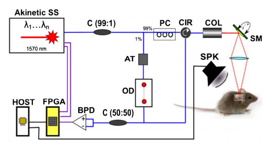
Fig. 1. Schematic of phase-sensitive OCT: SS, swept source; C (A:B), fiber coupler with A to B coupling ratio; AT, attenuator; OD, optical delay line; PC, polarization controller; CIR, circulator; COL, collimator; SM, scanning mirror; OL, objective lens; BPD, balanced photodetector; FPGA, FPGA module; HOST, host computer; SPK; speaker. Blue lines: optical connections. Purple lines: electrical SMA connections. Black lines: electrical BNC connections.
Doi: 10.1364/BOE.5.003547
Published: 2014.09.11
内容介绍:
Susana Marcos等人实现了利用OCT对于人眼晶体量化的三维表面形貌成像,研究了晶体前表面和后表面的表面面型的关系,以及它们随着年龄的变化。先对于由屈光介质的折射现象和折射率等影响造成的图像失真进行重构,再通过二维和三维的校正。主要包括对于不同表面的三维光线追迹、去噪、分割和扇形失真的校正,并借助6阶Zernike多项式逼近,从而大大提高对于曲率半径估计的准确性,尤其是表面的非球面性。该技术可以应用于在体检测角膜表面的高度图、生物统计、镜头对准、在调节状态下动态检测晶体的曲率,以及在体检测晶体的表面形貌。图2显示了原始的OCT前后段晶体的图像,以及分段的三维晶体面型。
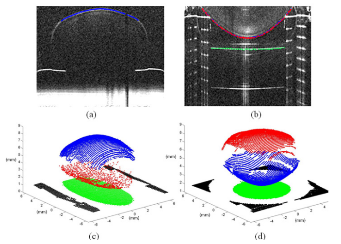
Fig. 2. Upper panels: Anterior-up OCT images corresponding to the anterior-up position of one of the crystalline lenses imaged, with (a) OCT focused on the anterior surface and (b) OCT focused on the posterior surface and the image of the cuvette. The detected surfaces are also marked in blue (anterior surface) red (posterior surface) and green (cuvette). Lower panels: 3-D OCT data from images of the crystalline lens with (c) the anterior surface up and (d) the posterior surface up. The blue and red points correspond to the segmented anterior and posterior surfaces of the lens, respectively. The green points and the black points correspond to the cuvette imaged through the lens, therefore distorted by the lens, and the cuvette without distortion respectively
试验中发现象散现象是造成晶体前后表面成像畸形的主要因素,其次是球差、慧差等因素的影响,具体所占的影响比重如下所示:
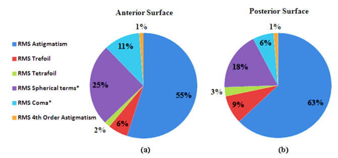
Fig. 3. Relative contribution of different Zernike terms to the overall surface elevation maps (in terms of RMS2) with an asterisk the terms that change statistically significantly with age.
二、 简讯:
Doi: 10.1364/OL.39.000041
Published: 2014.06.01
Abstract:
We report on a noncontact low-coherence optical phase-based imaging method, termed shear wave imaging optical coherence tomography (SWI-OCT), which enables 2D depth-resolved visualization of the low-amplitude elastic wave propagation in tissue with ultrahigh frame rate. SWI-OCT is based on 1D transverse scanning of the M-mode OCT imaging that is precisely synchronized with a low-pressure short-duration air-puff loading system. This approach of scanning and data recording allows visualization of the induced tissue deformation at high frame rate. The applied phase-resolved interferometric technique, with sensitivity on the nanometer scale, makes the low-amplitude tissue displacement detectable. For the demonstration of this method, and to study its application for tissue biomechanics, we performed pilot experiments on agar phantoms and ex vivo rabbit corneas. Samples with different elastic properties can be differentiated based on the velocity of the elastic wave propagation that is directly visualized with a 25 kHz frame rate. Our results indicate that SWI-OCT has the potential to be further developed as a major technique for depth-resolved high-resolution tissue elastography in vivo.
Doi: 10.1117/1.JBO.19.2.021107
Published: 2014.02
Abstract:
An approach to elastographic mapping in optical coherence tomography (OCT) using comparison of correlation stability of sequentially obtained intensity OCT images of the studied strained tissue is discussed. The basic idea is that for stiffer regions, the OCT image is distorted to a smaller degree. Consequently, cross-correlation maps obtained with compensation of trivial translational motion of the image parts using a sliding correlation window can represent the spatial distribution of the relative tissue stiffness. An important advantage of the proposed approach is that it allows one to avoid the stage of local-strain reconstruction via error-sensitive numerical differentiation of experimentally determined displacements. Another advantage is that the correlation stability (CS) approach intrinsically implies that for deformed softer tissue regions, cross-correlation should already be strongly decreased in contrast to the approaches based on initial reconstruction of displacements. This feature determines a much wider strain range of operability than the proposed approach and is favorable for its free-hand implementation using the OCT probe itself to deform the tissue. The CS approach can be implemented using either the image elements reflecting morphological structure of the tissue or performing the speckle-level cross-correlation. Examples of numerical simulations and experimental demonstrations using both phantom samples and in vivo obtained OCT images are presented.
Doi: 10.1364/OL.39.002888
Published 2014.05.12
Abstract:
We present an optofluidic optical coherence tomography (OCT) needle probe capable of modifying the local optical properties of tissue to improve needle-probe imaging performance. The side-viewing probe comprises an all-fiber-optic design encased in a hypodermic needle (outer diameter 720 mu m) and integrates a coaxial fluid-filled channel, terminated by an outlet adjacent to the imaging window, allowing focal injection of fluid to a target tissue. This is the first fully integrated OCT needle probe design to incorporate fluid injection into the imaging mechanism. The utility of this probe is demonstrated in air-filled sheep lungs, where injection of small quantities of saline is shown, by local refractive index matching, to greatly improve image penetration through multiple layers of alveoli. 3D OCT images are correlated against histology, showing improvement in the capability to image lung structures such as bronchioles and blood vessels. (C) 2014 Optical Society of America.
Doi 10.1364/OE.22.007514
Published:2014.04.07
Abstract:
We report the enhancement in the obtained signal and penetration depth of 2-D depth-resolved images that were taken by shaping the incident wavefront in optical coherence tomography (OCT). Limitations in the penetration depth and signal to noise ratio (SNR) in OCT are mainly due to multiple scattering, which have been effectively suppressed by controlling the incident wavefront using a digital mirror device (DMD) in combination with spectral-domain OCT. The successful enhancements in the penetration depth and SNR are demonstrated in a wide-range of tissue phantoms, reaching depth enhancement of up to 92%. The hidden structures inside a tissue phantom that could not be seen in conventional OCT are clearly revealed through our proposed system. Its 2-D imaging capability, assisted by further optimization of the system for real-time acquisition speed will boost wide-spread use of OCT for in-vivo tissue diagnosis. (C) 2014 Optical Society of America
供稿:倪秧