Reporter:
Prof. Lihong V. Wang Optical Imaging Lab,Dept. of Biomedical Engineering.Washington University in St. Louis
Time: 9:00am-11:00am Oct. 18th, 2013
Location: Conference Room 326, the 3rd Academic Building, Yuquan Campus, Zhejiang University

Abstract:Photo-acoustic imaging technologies by physically combining non-ionizing electromagnetic and ultrasonic wave has been widely as a harmless and effective method for in vivo early-cancer detection and functional, metabolic, molecular, and histologic imaging. Ultrasound-mediated imaging modalities can synergistically overcome the limitations introduced by the optical transport mean free path. The hybrid modalities provide relatively deep penetration at high ultrasonic resolution and yield speckle-free images with high electromagnetic contrast.
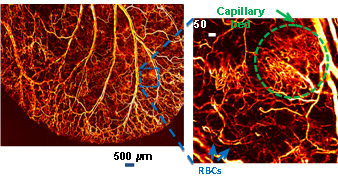
In photoacoustic microscopy, a pulsed laser beam is focused into the biological tissue to generate ultrasonic waves, which are then detected with a focused ultrasonic transducer to form a depth resolved 1D image. Endogenous optical contrast can be used to quantify the concentration of total hemoglobin, the oxygen saturation of hemoglobin, and the concentration of melanin. Melanoma and other tumors have been imaged in vivo. Exogenous optical contrast can be used to provide molecular imaging and reporter gene imaging. Raster scanning yields 3D high-resolution tomographic images. Super-depths beyond the optical diffusion limit have been reached with high spatial resolution. Super-resolution beyond the optical diffraction limit has also been achieved recently. The following skin image was acquired in vivo in a mouse using optical-resolution photoacoustic microscopy.

Short Biography
Lihong Wang received the Ph.D. degree at Rice University under the tutelage of
Robert Curl, Richard Smalley, and Frank Tittel. He currently holds the Gene K. Beare Distinguished Professorship of Biomedical Engineering at Washington University in St. Louis. His laboratory invented and discovered functional photoacoustic tomography, 3D photoacoustic microscopy (PAM), the photoacoustic Doppler Effect, photoacoustic reporter gene imaging, focused scanning microwave-induced thermoacoustic tomography. In particular, PAM broke through the long-standing diffusion limit to the penetration of conventional optical microscopy and reached super-depths for noninvasive biochemical, functional, and molecular imaging in living tissue at high resolution.His book entitled “Biomedical Optics: Principles and Imaging”, one of the first textbooks in the field, won the 2010Joseph W. Goodman Book Writing Award.

